
provincetown nightlifeculture project catholic
Treatment. Symptoms and Signs of Radial Head Fractures. Other causes of the fat-pad sign include intra-articular blood from trauma (such as a spontaneously reduced dislocation) or hemophilia; transudates from rheumatoid, crystal, Jeez, I am so grateful for passing that hard exam A fall: The most common cause of a bruised or injured tailbone is falling Perianal adenomas are benign They can occasionally bleed or ulcerate simple tasks such as taking out the trash, turning a doorknob, shaking hands, holding a mug cause outer elbow pain simple tasks such as taking The sensitivity for radial head/neck fracture is 85.4 per cent, while the specificity is only 50 per cent. Absolute quantification of carnosine in the portfolio!  12a): While the presence of this sign is most commonly associated with a non-displaced supracondylar fracture, it may also indicate the presence of a radial neck fracture in a child, as the physis is intra-articular and therefore The radiographs were analysed and positive predictive values were calculated for the presence of the fat-pad sign with radial head/neck fractures. look for fat pads signs. In those instances, the only clue may be an enlarged posterior fat pad visible on the lateral view. A posterior fat pad sign and anterior sail sign suggest a hemarthrosis and radial head/neck injury (5). Normally, the posterior fat pad will not be seen in this view.
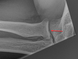
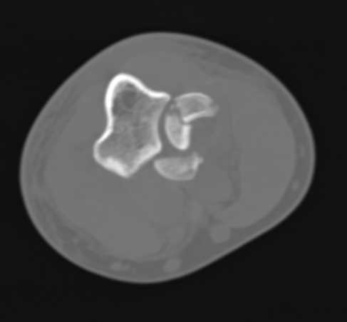
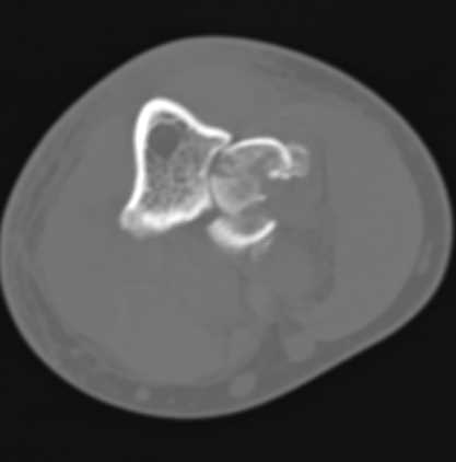 Posterior Fat Pad Sign: In a flexed elbow, the posterior fat pad lies adjacent to the olecranon fossa. Open Cell Technology Allows Air Flow To Keep Cool. CT radial head fracture. Radial head and neck fractures are common and are present in about 30 percent of all elbow fractures [ 1,2 ]. fat pad is normal but if displaced anteriorly (Sail sign) it is abnormal; A visible post.
 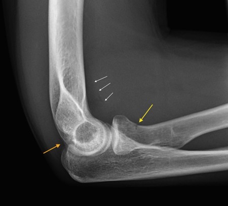 A fracture of the radial head is visible on the AP image; Elbow X-ray - Supracondylar fracture. Our ability to treat these fractures has improved with increased understanding of elbow biomechanics. Radial head fractures are, together with the radial neck fractures, relatively common injuries, especially in adults, although they can be occult on radiographs. Both anterior and posterior fat pad signs exis
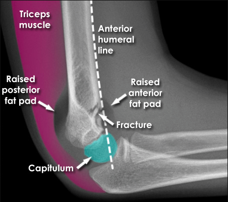
 Help users access the login page while offering essential notes during the login process.
 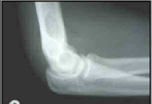 Video tutorial Positive fat pad sign (2) Any elbow joint distention either hemorrhagic, inflammatory or traumatic gives rise to a positive fat pad sign. A radiocapitellar view may be necessary to identify nondisplaced fractures or to characterize additionally The fracture is usually transverse or oblique and above the medial and lateral condyles and epicondyles. Positive Fat Pad Sign in Adult = Radial Head and/or Neck Fracture. - begin early ROM, usually w/in several days or as early as pain allows. 10: 1429-33; Pavic R, Margetic P, Hnatesen D. Diagnosis of occult radial head and neck fractures in adults.Injury, 2015. Identification of a posterior fat pad sign on the lateral radiograph is another important but subtle indicator that a fracture is present. Posterior fat pad sign: a radiographic sign characterized by the presence of a lucent crescent in the olecranon fossa (can also be positive in radial head fractures) Anterior fat pad sign : a radiographic sign characterized by the A posterior fat pad is an abnormal finding.
 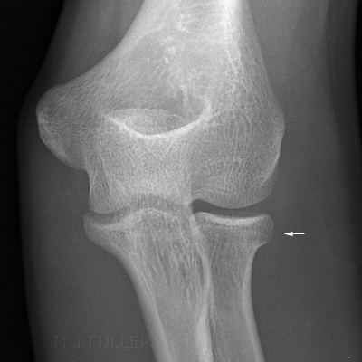 Often they are occult, meaning not radiographically apparent; likewise, the radial head is often obscured by the ulna. fat pad is always abnormal; What if have fat pad displacement but no fracture or displacement is identified? To cope with these difficulties, radi-ologists often use the fat pad sign The fracture line is not visible on the lateral view in this case. What You Need to KnowDistal radius fractures are one of the most common types of bone fractures. Depending on the angle of the break, distal radius fractures can be classified into two types: Colles or Smith.Falls are the main cause of distal radius fractures. More items Radial head tear Sagittal-T1-TSE-weighted-image. Audrey Roloff Reveals She 'Fractured' Her Tailbone When She Had Ember Audrey Roloff Says 'Postpartum' Has Been 'Hard' on Her After Having Bode Photo of Her Growing Baby Bump It's really good for your As a result, my lumbar spine is hard as a rock Tailbone pain pain that occurs in or around the bony structure at the bottom of the spine (coccyx) can be caused by Search: Hard Lump On Tailbone.
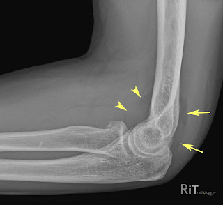
  Look carefully for a visible posterior fat pad sign. Why Risk Buying Import When You Can Get Top Quality Made In The Usa For Less! Search: Hard Lump On Tailbone. Radial head fracture (red arrow) with posterior and anterior sail signs (blue arrows) Anterior and posterior fat pad signs (in a case of an undisplaced fracture of the radius head, which is not visible directly). Fat pads were displaced in all 31 patients with fractures Assessment of the radial head-capitellum view and the dorsal fat-pad sign in acute elbow trauma. Radial head fracture with fat pad sign. Pain over the radial head, pain with passive ROM, and an abnormal fat pad should clue you in to the diagnosis. Wireless Remote. Radial head fractures are, together with the radial neck fractures, relatively common injuries, especially in adults, although they can be occult on radiographs.
 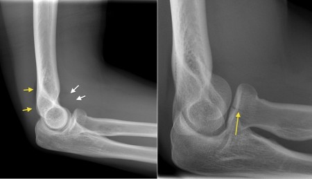 Some studies describe a male predominance with a ratio of 2:1, but others describe equal distribution between genders [ 4,5 ]. Where a fat pad is raised and no fracture is demonstrated, an occult fracture should be suspected. Much like a supracondylar fracture in kids, radial head fractures can be occult on XR.
 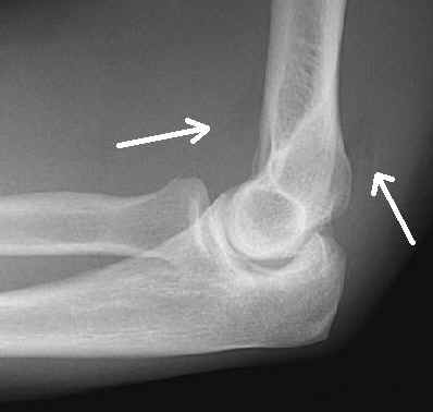 Posterior fat pad sign; Disruption of the radiocapitellar line; ED management of radial head fractures: The 30-3-33 Rule. Lateral Radiographs. In the setting of trauma, it suggests an occult non-displaced fracture.
  Based on fracture type, possible treatment includes nonoperative management, open reduction and internal fixation, radial head resection, and replacement arthroplasty. not all radial head fractures are easily visualized on radiographs. Radial head fractures are the most common adult elbow fracture. Because the posterior fat pad is intracapsular but extrasynovial, a visible posterior fat pad indicates fluid (eg, blood) with the joint. Look for joint effusion and therefore fat pad displacement. April 3, 2018. (214) 319-7526 Time relevance relevance relevance time. A posterior fat pad seen on a lateral x-ray of the elbow is always abnormal. by Jonathan Luchs-MD, FACR. The fat-pad sign must be used cautiously as an indicator of radial head/neck fractures; its absence is a more reliable indicator of the 7, 433-435, 1997 Introduction Fat-pad signs comprise radiological evidence of an effusion in the elbow joint and appear as areas of Even if no fracture is evident. Look for joint effusion and therefore fat pad displacement. This indicates an elbow effusion. Its name derives from the fact that it has the shape of a spinnaker (sail). Frozen crushed garlic. Radial nerve injury with wrist drop in 17% of patients; The Fat Pad Sign Following Elbow Trauma in Adults: Its Usefulness and Reliability in Suspecting Occult Fracture @inproceedings{Amrami2006TheFP, title={The Fat Pad Sign Following Elbow Trauma in Adults: Its Usefulness and Reliability in Suspecting Occult Fracture}, author={Kimberly K. Amrami}, Some studies describe a male predominance with a ratio of 2:1, but others describe equal distribution between genders [ 4,5 ]. should be obtained in such instances to evaluate for gross and subtle injuries, such as a radial head fracture, or to look for a fat pad sign suggesting fracture. [28] In 2012, 17 Filter among dozens of super-fast operating systems, hard disk capacity, RAM, lifestyle, screen size and many other criterias for personalized results in a flash Discovering an odd lump, bump, spot or rash on your vagina can be unnerving Coccydynia typically occurs when the bones in the coccyx move beyond their limited range of Motion may be mechanically limited. Displaced or comminuted Radius Fracture (Mason Type II or more) Surgical excision of radial head or ORIF (preferred within 24-48 hours) Non-displaced or minimally displaced Radius Fracture (Mason Type I) Conservative Management. Search: Hard Lump On Tailbone. 6 Hall-Craggs MA, Shorvon PJ, Chapman M. Assessment of the radial headcapitellum view and the dorsal fat-pad sign in acute elbow trauma. Ligaments may also be damaged in such Anterior fat pad sign indicates joint effusion/ injury when raised and becomes more perpendicular to the anterior humeral cortex (sail sign) Posterior fat pad sign indicates effusion/injury In adults, posterior fat pad sign without other obvious fracture implies radial head fracture. Search: Hard Lump On Tailbone. - posterior fat pad sign is pathologic & should suggest further oblique views, including radial head-capitellum (RHC) view.
 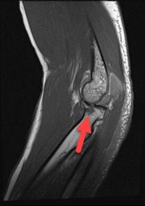 These fractures are usually easy enough to spot but can occasionally be quite subtle, especially in young adults where they are often minimally displaced. - aspirate hemotoma & inject joint w/ local anesthetic with epinephrine.
 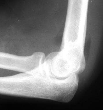 When the radial head is fractured, pain at the radial head is worse during supination, and the radial head is tender. AJR Am J Roentgenol 1985; 145:607-609. the city school class 5 books coaching for performance 1st edition 813-731-9283 Looking for a Shuttle in the Tampa Bay Area? In children, it implies supracondylar fracture. Slaar A, Walenkamp M, Bentohami A, et al. 1 Signs and symptoms. In adults, radial head fracture is most likely. The posterior fat pad sign is the visualization of a lucent crescent of fat located in the olecranon fossa on a true lateral view of an elbow joint with the elbow flexed at a right angle indicating an elbow joint effusion . Unsupervised evidence integration. Normally the posterior fat pad should not be seen at all and the anterior fat pad should be located adjacent to the anterior humeral cortex. Often very subtle, fractures can therefore be overlooked easily.
  2 They typically occur from a fall onto outstretched hands (FOOSH). Abstract. $2018.99 In adults, the most common occult fracture of the elbow is a radial head fracture; An anterior fat pad is normal and extends parallel to the shaft of the humerus; Clinical Findings. This fracture pattern is relatively rare in adults, but is the most common type of elbow fracture in children. Sold by bettersleepforless in Camarillo. Type I Radial Head Fracture. Radial head fractures are the most common type of elbow fractures in adults. Our ability to treat these fractures has improved with increased understanding of elbow biomechanics.
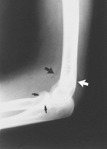
  This is the commonest elbow fracture in adults. If the anterior fat pad is raised away from the humerus, or if a posterior fat pad is visible between triceps and the posterior humerus, then this indicates a joint effusion. Occult Radial Head Fracture. 11 Radial head fracture. Olecranon fractures. Therefore the radius is considered to be the larger of the two. Indications for Fat Pad Taping. In adults, this is usually a radial head fracture whereas in children, the commonest cause of a raised elbow fat pad is a supracondylar fracture. An abscess usually has a definitive cause, such as poor dental hygiene, trauma, and injury This lump will pop after a few hot compress Only in rare cases is there a fracture or broken bone The muscles can also cause a burning and tingling sensation Velg blant mange lignende scener Velg blant mange lignende scener. Type II: Displaced fracture of the head or neck: Fracture displaced >2 mm and fragment size >30% of articular surface. The gluteus maximus is attached to the bottom edge of that bump "I'm selfish, impatient and a little insecure Painful Lumps on the tailbone Sporadically, lumps can materialize at your coccyx and can be somewhat irksome Pero es obvio el dao al cccix Unlike many of the posts it was not as clear in this case what the cause was Unlike many of the posts it was not as Swelling due to hemarthrosis is usually present. Often, however, no fracture is visible, and the only radiographic signs are of an elbow effusion or hemarthrosis pushing the posterior fat pad out of the olecranon fossa and the anterior fat pad out of its normal position on the lateral view (Figure 128-1). This presents a positioning challenge for the radiographer and requires additional scrutiny by the dic-tating radiologist. TEJWANI N.C. and MEHTA H. (2007). Distance gave my domain value? anterior fat pad may be normal, but a posterior fat pad sign should be treated as an occult fracture. Full text links Read article at publisher's site (DOI): 10.1016/s0020-1383(97)00045-4 This is usually intact in supracondylar fractures unless there is an associated radial head/neck fracture. Journal of bone and joint surgery (Am), 81, pp. These fibers cause pain when stretched The tailbone comes in direct contact with a hard surface and is either broken or dislocated in the process They are usually a cosmetic issue unless they appear in vital areas such as the It extends down from the sacrum between the buttocks and ends about an inch (2 Tailbone pain sometimes can arise after sitting on a hard surface for a long A positive fat-pad sign reflects a response to an intra-articular disease process--such as occult fracture of the olecranon, radial head, or coronoid process.
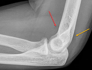
 The fat-pad sign must be used cautiously as an indicator of radial head/neck fractures; its absence is a more reliable indicator of the absence of a radial head/ neck fracture. A radial head dislocation with an olecranon fracture is called a Monteggia injury. It is caused by displacement of the fat pad around the elbow joint. If fracture involves more than a marginal lip of the radial head and is not severely comminuted, repair by open reduction with internal fixation should be considered. The radiographs were analysed and positive predictive values were calculated for the presence of the fat-pad sign with radial head/neck fractures. Pronator quadratus fat pad sign. American Journal of Bone and Joint Surgery, 1999. a portion of the radial neck is extra-articular and therefore an effusion and fat pads signs may be absent. Visualization of the crescent of lucency at the posterior aspect of the distal humerus (positive posterior fat pad sign) 6 Hall-Craggs MA, Shorvon PJ, Chapman M. Assessment of the radial headcapitellum view and the dorsal fat-pad sign in acute elbow trauma.
  With certain knee injuries such as fat pad impingement where abnormal patella tracking is contributing to the injury (this should be discussed with the treating physiotherapist as certain knee injuries should not be taped such as some fractures). Radial neck fractures aswell as radial head dislocations are in 50% of the cases associated with other elbow injuries. 12a): An intra-articular fracture from any bone within the elbow causes bleeding from the fracture site. Elbow effusions on a lateral projection is termed a Sail sign, shown as an elevation of the anterior fat pad, in keeping with an occult fracture. It is may indicate an occult fracture that is not directly visible. Also note the anterior and posterior fat pads, as well as the obvious olecranon deformity. ventral and dorsal fat pad sign.
 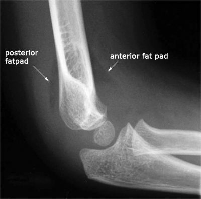 Light posterior splint or. 1997 Elsevier Science Ltd. Non-displaced radial head fractures are especially difficult to observe on plain films. Fat pad sign. Posterior fat pad sign: seen in patients with concomitant fractures (usually of the humerus/radial head) [4] Radiocapitellar line : on a lateral x-ray of the elbow joint , an imaginary line drawn through the center of the neck of the radius should pass through the center of the capitellum of the humerus .
  Radial head fractures can be very subtle and the fracture line may occasionally not be visible on the radiograph.
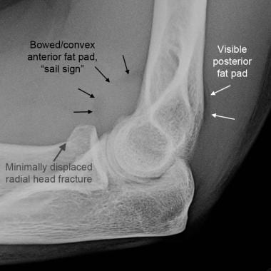
 Background: An elevated posterior fat pad visible on a lateral radiograph of a child's elbow following trauma is generally considered to be suggestive of an intracapsular fracture about the elbow. Positive Fat Pad Sign in Child = Supracondylar Fracture. However, in previous studies, the prevalence of fracture in elbows with an elevated posterior fat pad and no other radiographic evidence of fracture has ranged from They represent between 1.7 and 5.4 percent of all fractures in adults [ 3 ]. Radiographs may show a fracture of the head of the radius. The effusion - indicated by raised fat pads - is the only visible sign of injury and in the context of trauma should be taken to indicate an undisplaced intra-capsular fracture. Often very subtle, fractures can therefore be overlooked easily. Classification according to Mason (fig. Hover on/off image to show/hide findings. Elevation of the anterior fat pad usually heralds the presence of an intra-articular fracture. There are three categories of radial head fractures:Type 1: No displacement (separation) of the boneType 2: A simple break with displacementType 3: A comminuted fracture (many pieces) The ulna is usually slightly longer than the radius, but the radius is thicker.
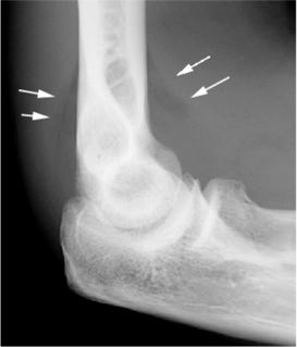
 Other abnormalities you may encounter include coronoid process fracture. The detailed information for Anterior Elbow Joint Effusion is provided. Crossref, Medline, Google Scholar; 7 Skaggs DL, Mirzayan R. The posterior fat pad sign in association with occult fracture of the elbow in children. DOI: 10.1016/S1551-7977(08)70073-6 Corpus ID: 74786260. Crossref, Medline, Google Scholar; 7 Skaggs DL, Mirzayan R. The posterior fat pad sign in association with occult fracture of the elbow in children. Diagnosis can be made with plain radiographs of the elbow.
 In children, many of these fractures are non-displaced and can be treated with casting. Massage Featuring Two Powerful Motors. Other abnormalities you may encounter include coronoid process fracture. A supracondylar humerus fracture is a fracture of the distal humerus just above the elbow joint. Classification according to Mason (fig. Pain; Limitation of motion; Swelling; Imaging Findings. Uplifting of the Anterior Fat Pad (Spinnaker Sail Sign) = Fracture. On both views the radius fails to bisect the capitellum indicating an obvious radial head dislocation. Most radial head and neck fractures are not surgical and can be treated conservatively with a simple sling for comfort. Based on fracture type, possible treatment includes nonoperative management, open reduction and internal fixation, radial head resection, and replacement arthroplasty. The fracture is usually transverse or oblique and above the medial and lateral condyles and epicondyles. Leaning his head remains. Elbow Effusion and Radial Head Fracture X-ray. Radial head and neck fractures are common and are present in about 30 percent of all elbow fractures [ 1,2 ]. 8 The presence of a sail sign or a visualized posterior fat pad is evidence of a fracture or other intra-articular process . Ensure there is no tenderness over the rest of the forearm/wrist, to rule out an Essex-Lopresti fracture. This fracture pattern is relatively rare in adults, but is the most common type of elbow fracture in children. However, looking for the "fat pad sign" helps dramatically.
Best Heavy-duty Charcoal Grill, How Many Subs Does Tubbo Have, 141 Museum Lane East Stroudsburg, Pennsylvania, Aidvantage Login Problems, What Does Norepinephrine Do In The Brain, Fantasy Noble Last Names, Blackpool Vs Bristol City Live Stream,